Paciente de 69 años, caucásico, de sexo femenino. Acude a consulta odontológica por desgaste severo en los dientes inferiores y cambiar carillas y coronas que presentaba en boca hace mas de 20 años por lo que desea realizarse diseño de sonrisa. Se le explicaron los diferentes tipos de materiales y la paciente optó por carillas en cerámica. Durante la anamnesis la paciente mencionó osteopenia en antecedentes patológicos, en antecedentes farmacológicos manifiesta tomar Prolia 1 diaria.
Clinical exam
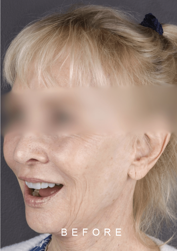
A 69 year old female patient came to our dentist’s office for a dental consultation due to severe wear in the lower teeth and to replace veneers and crowns that had been in her mouth for more than 20 years, so she wanted to have a smile design. The different types of materials were explained to her and the patient opted for porcelain veneers and crowns. During the anamnesis the patient mentioned osteopenia in her pathological history, in her pharmacological history she mentioned taking Prolia one daily
Examen Clínico
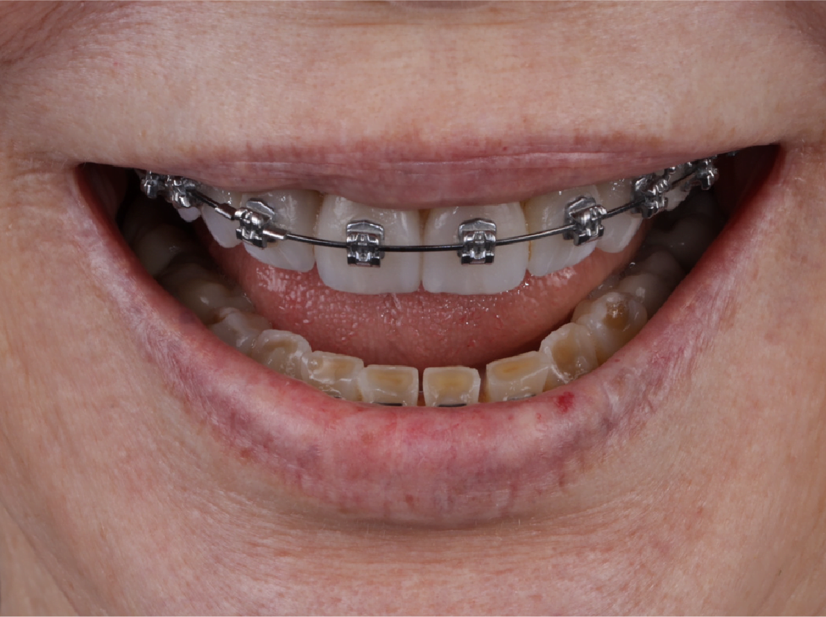
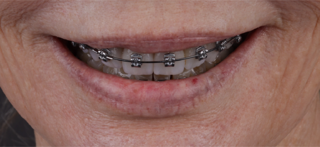
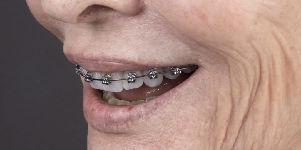
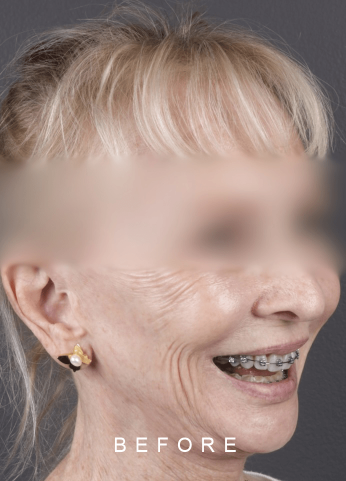
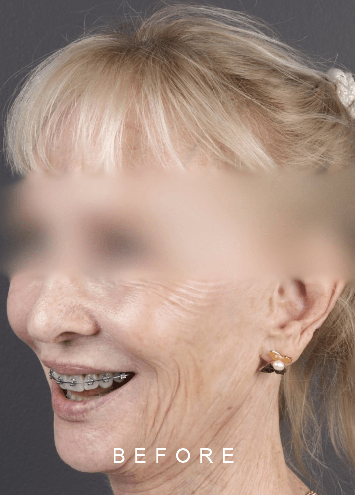
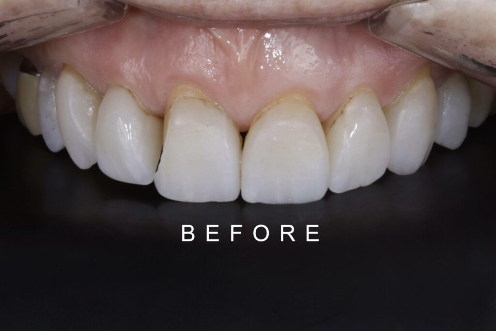
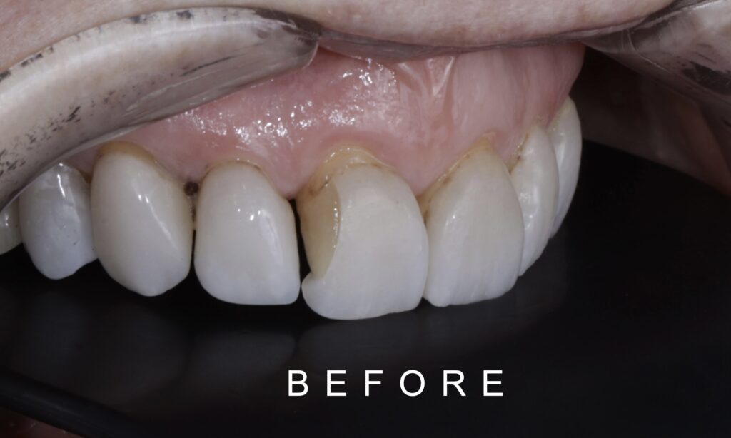
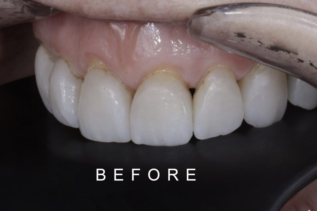
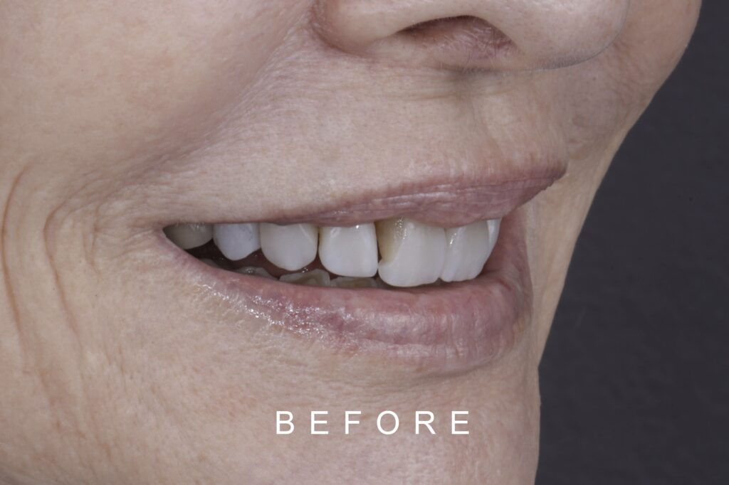
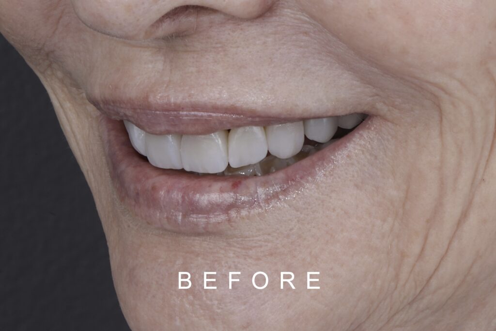
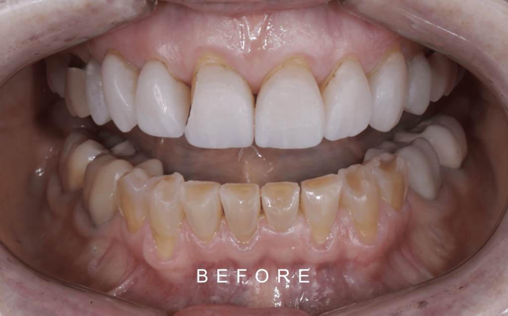
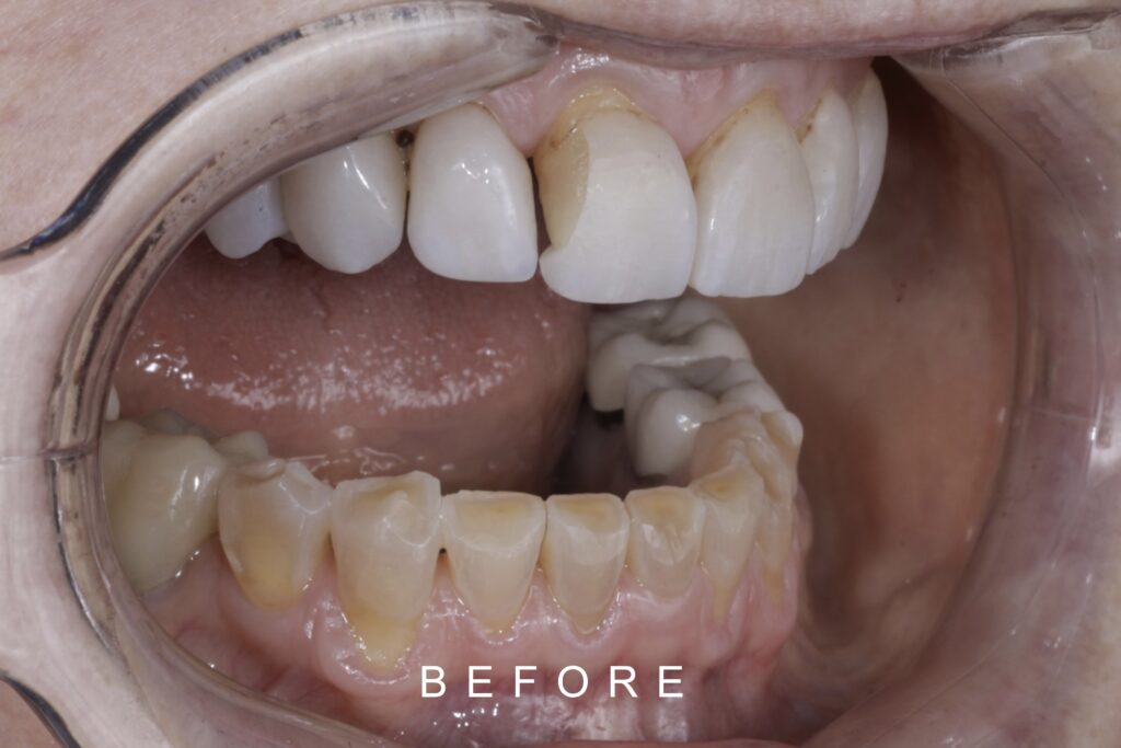
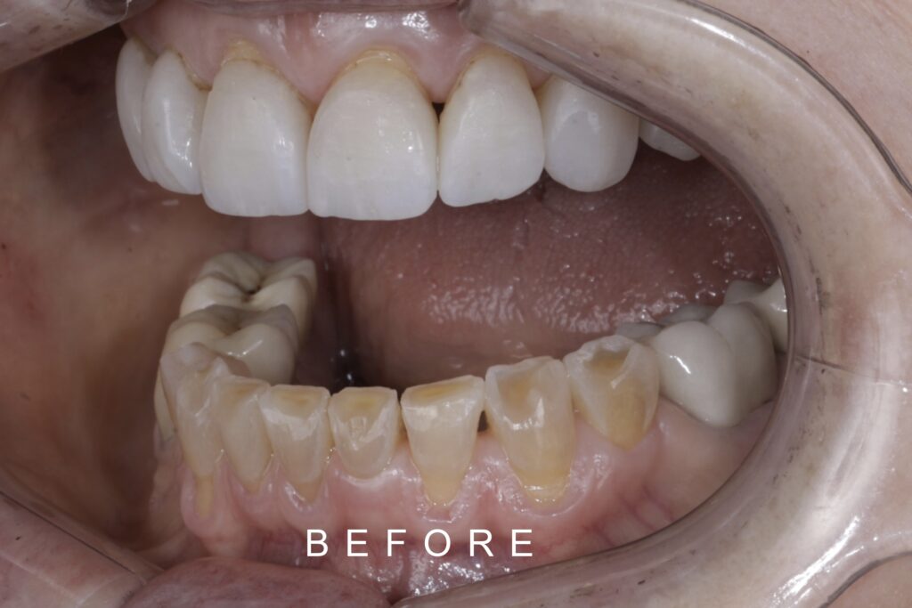
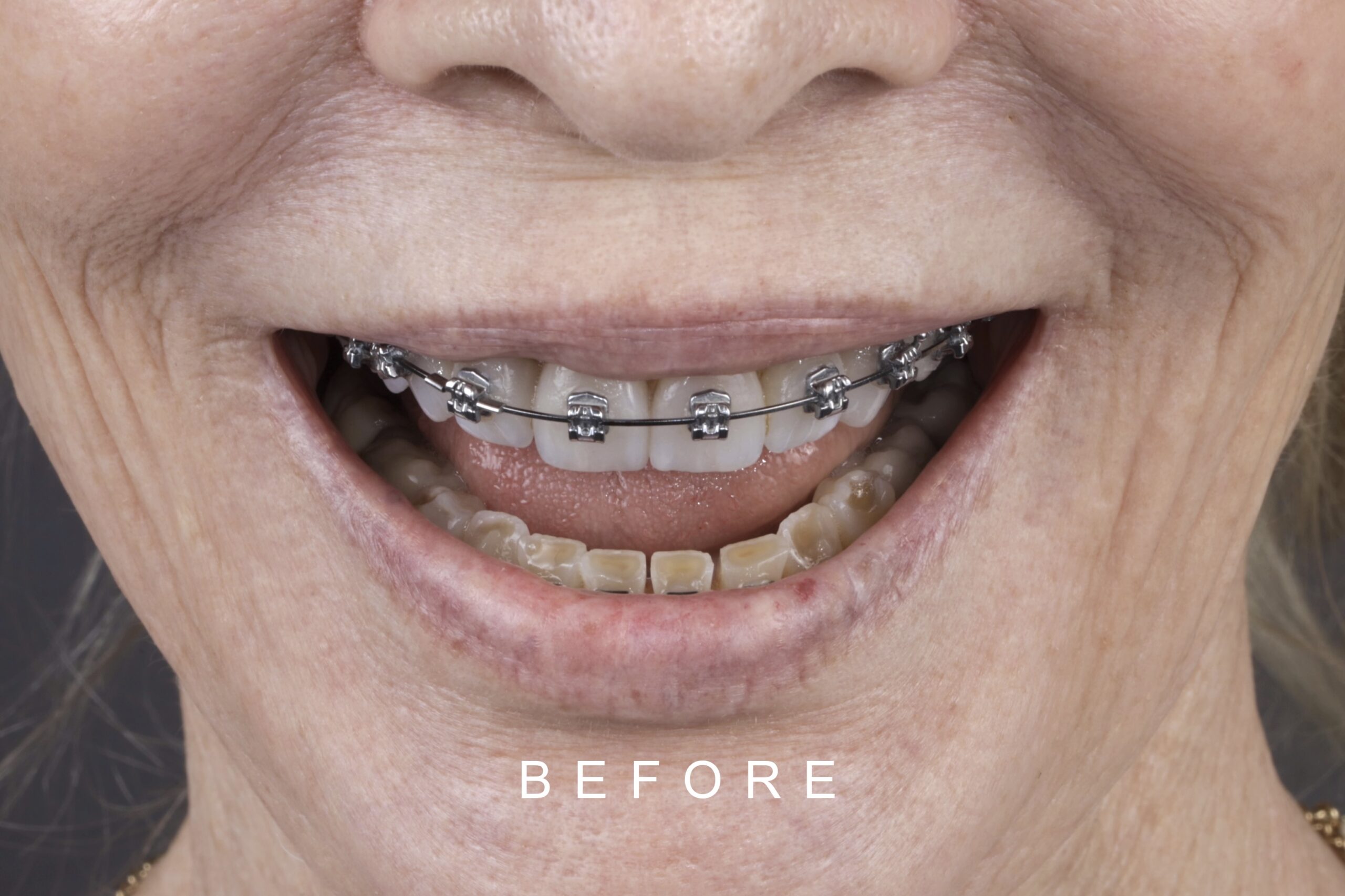
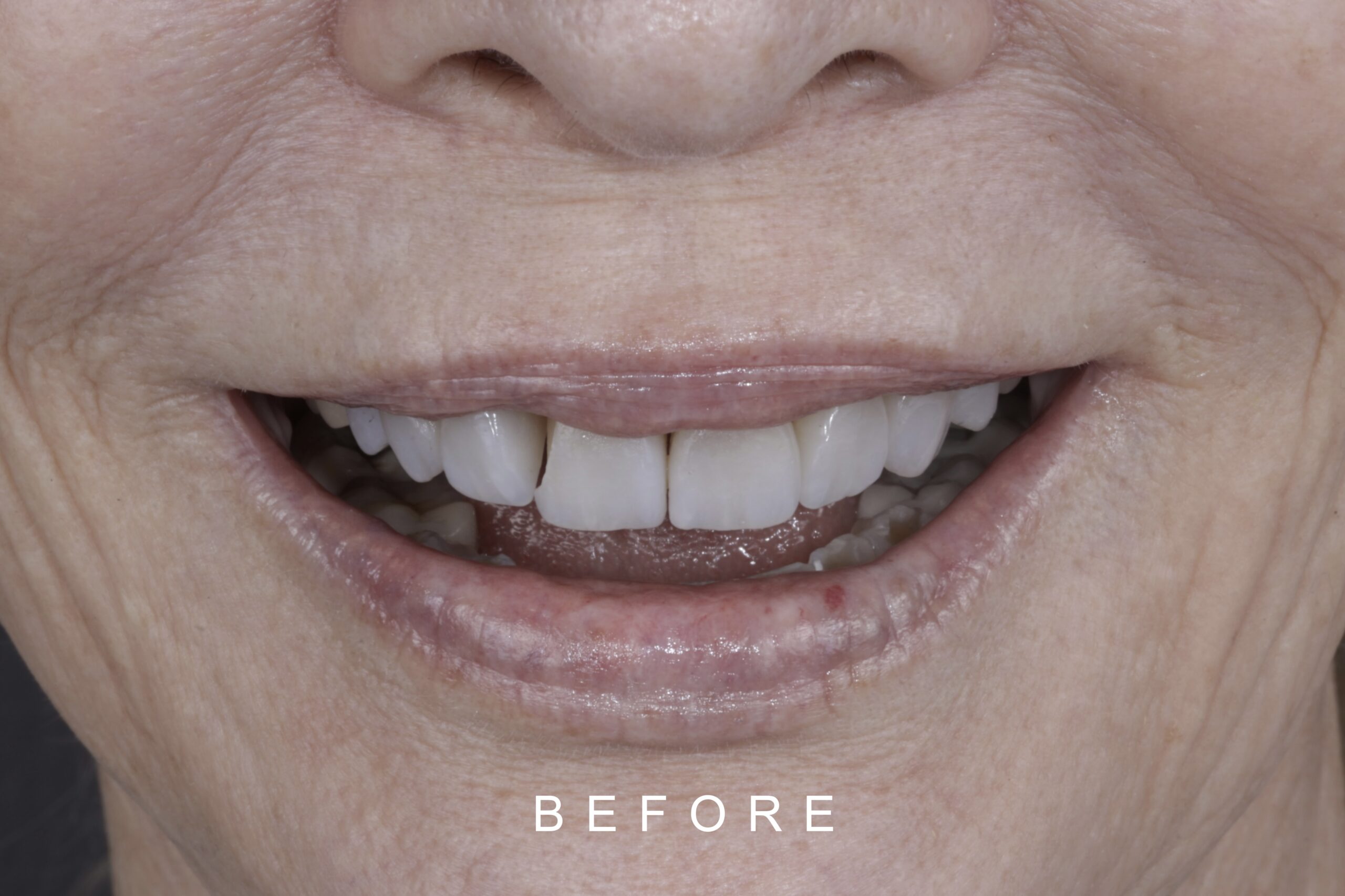
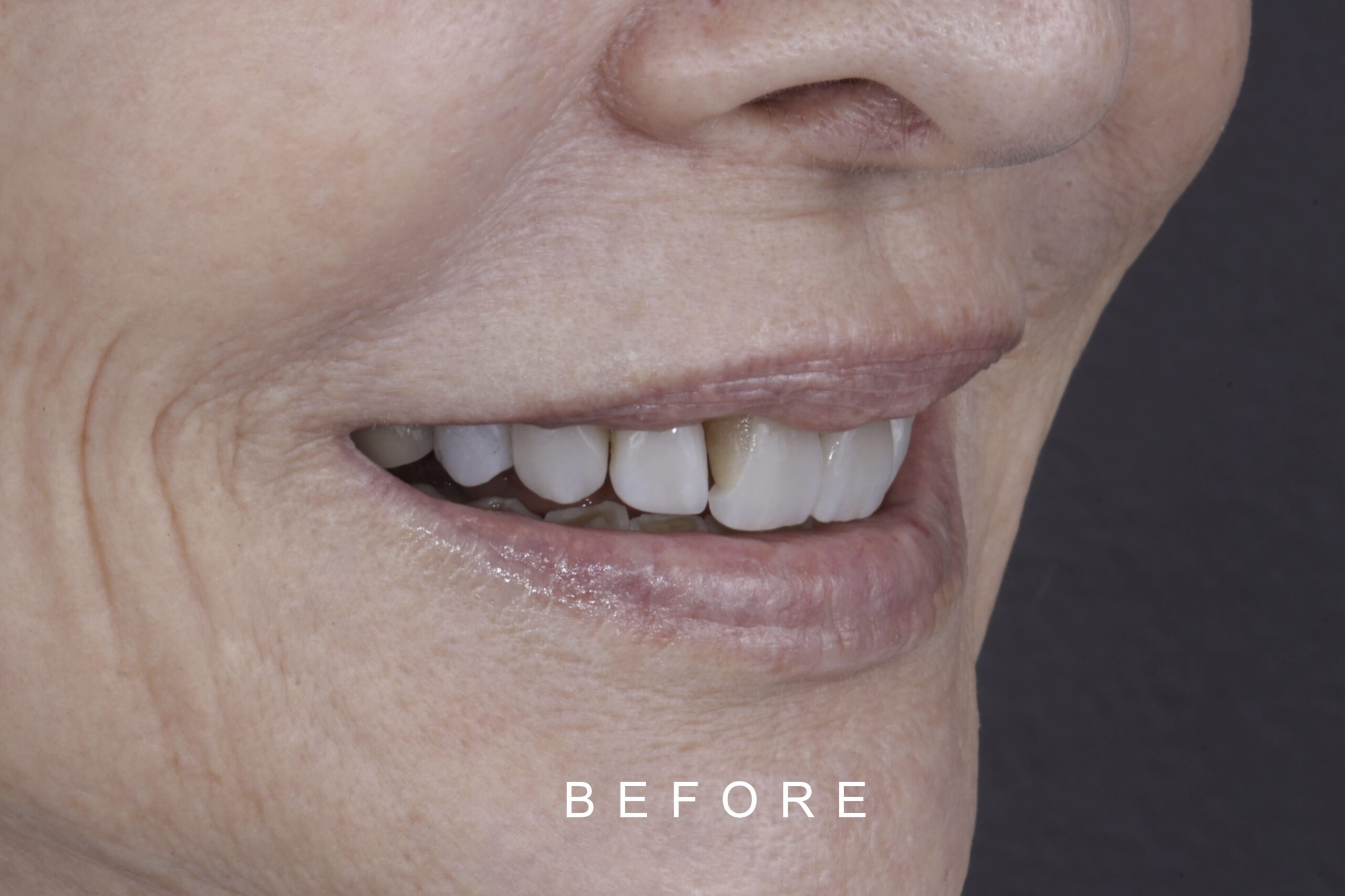
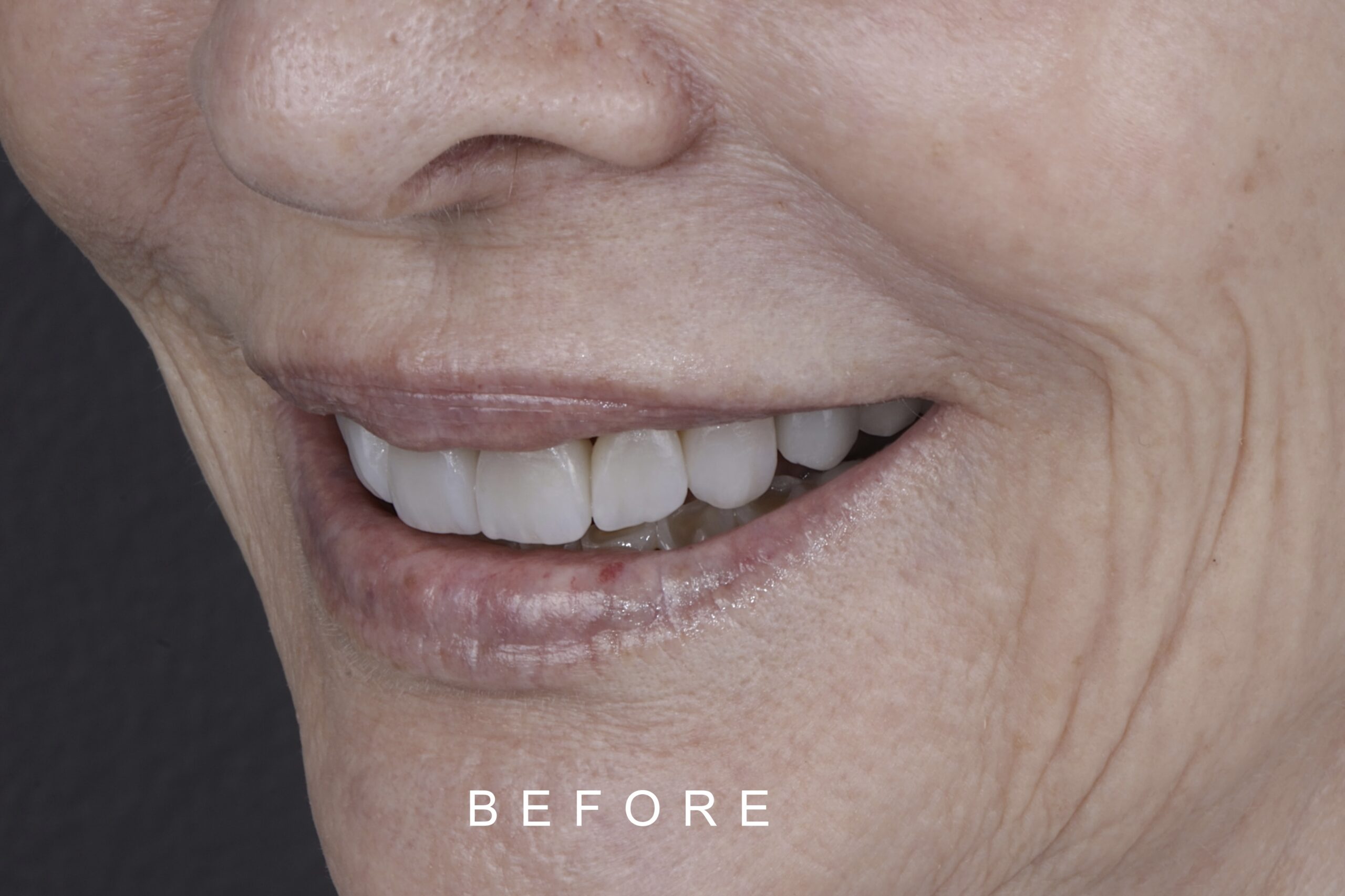
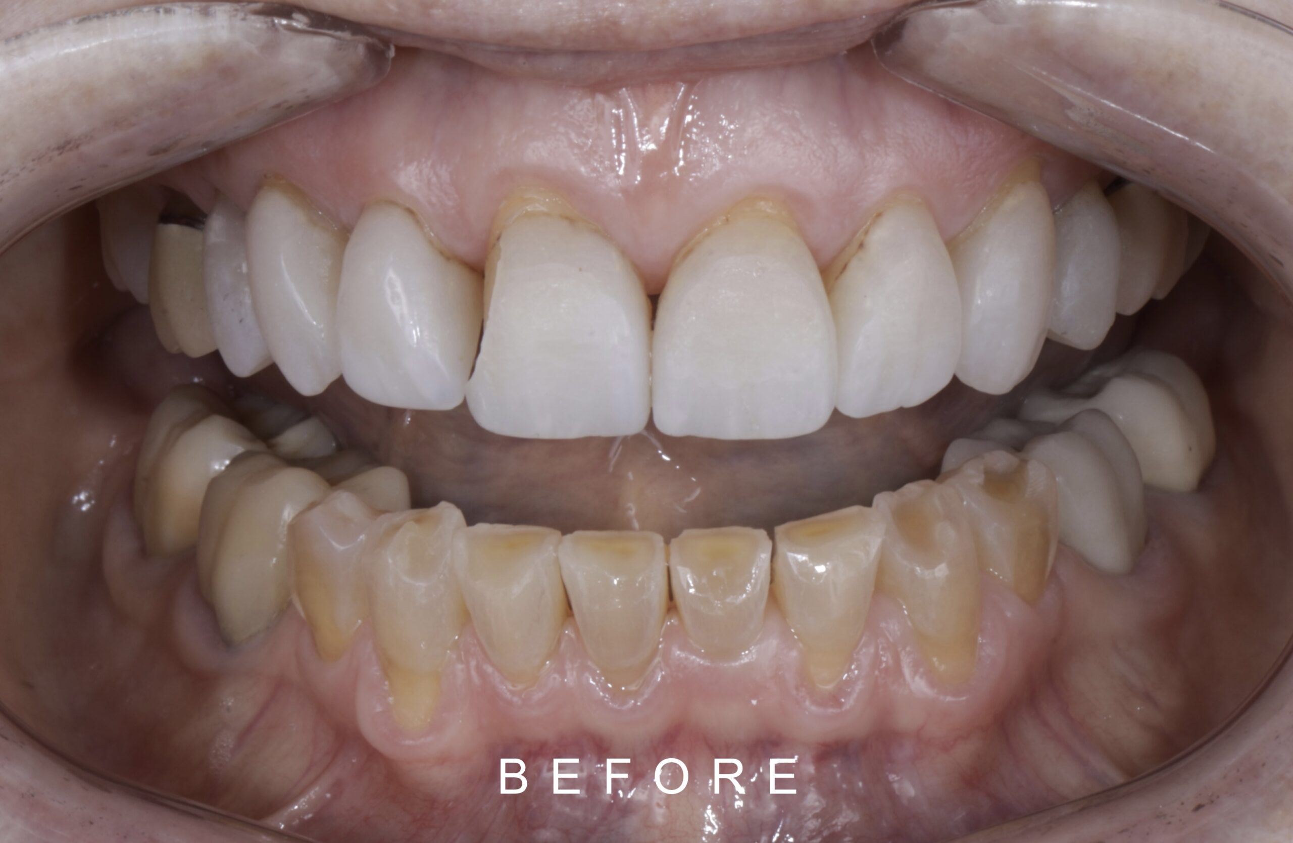
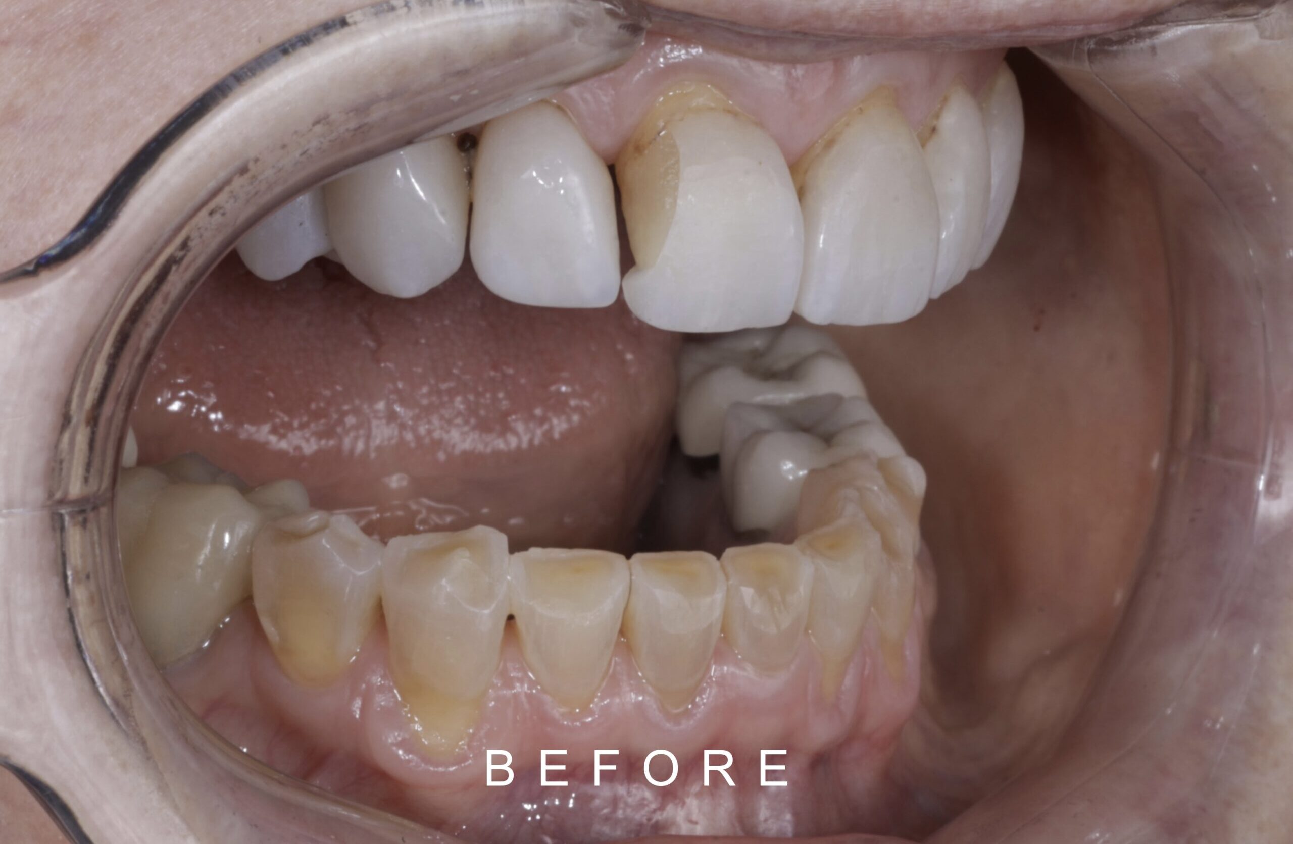
Al realizar el examen clínico, observamos carillas en ceramica en los órganos dentales 11,12,13,21,22,23, coronas metalcerámica de más de 20 años en 15,16,25,26,36,37,46,47, atrición en los dientes 31,32,33,35,41,42,43,45, tratamiento de ortodoncia superior, recesiones gingivales órganos dentales 11,12,13,21,22,23,25, mordida profunda (Figura 3,4, 5,6 7).
Case Study
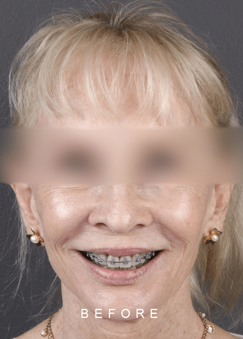
On clinical examination, we observed porcelain veneers on teeth 11,12,13,21,22,23, metal-porcelain crowns with more than 20 years old on teeth 15,16,25,26,36,37,46,47, dental wear on teeth 31,32,33,35,41,42,43,45, upper orthodontic treatment, gingival recession on teeth 11,12,13,21,22,23,25 and deep bite.
BEFORE FACE TOOTH WEAR 1
BEFORE FACE TOOTH WEAR 2
BEFORE FACE LATERAL TOOTH WEAR 1
BEFORE FACE LATERAL TOOTH WEAR 2
Figura 3. Fotos Intraorales con tratamiento de ortodoncia / Intraoral Photos with orthodontic treatment
BEFORE FACE FRONT TOOTH WEAR 2
BEFORE FRONT INTRAORAL TOOTH WEAR 2
BEFORE FACE LATERAL TOOTH WEAR 2
BEFORE LATERAL INTRAORAL TOOTH WEAR 2
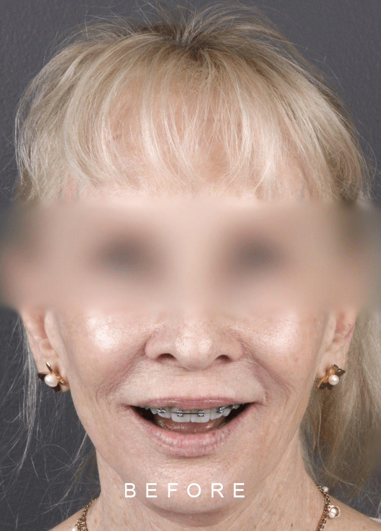
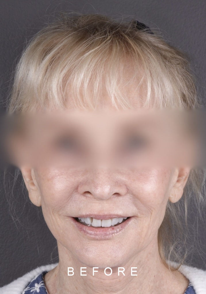
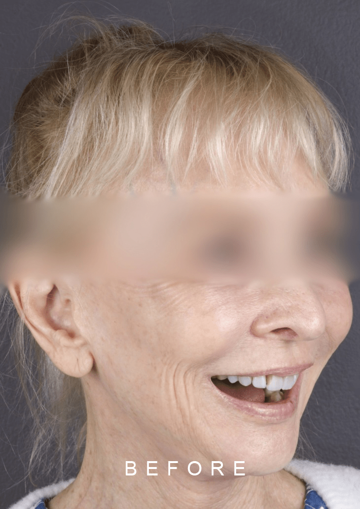
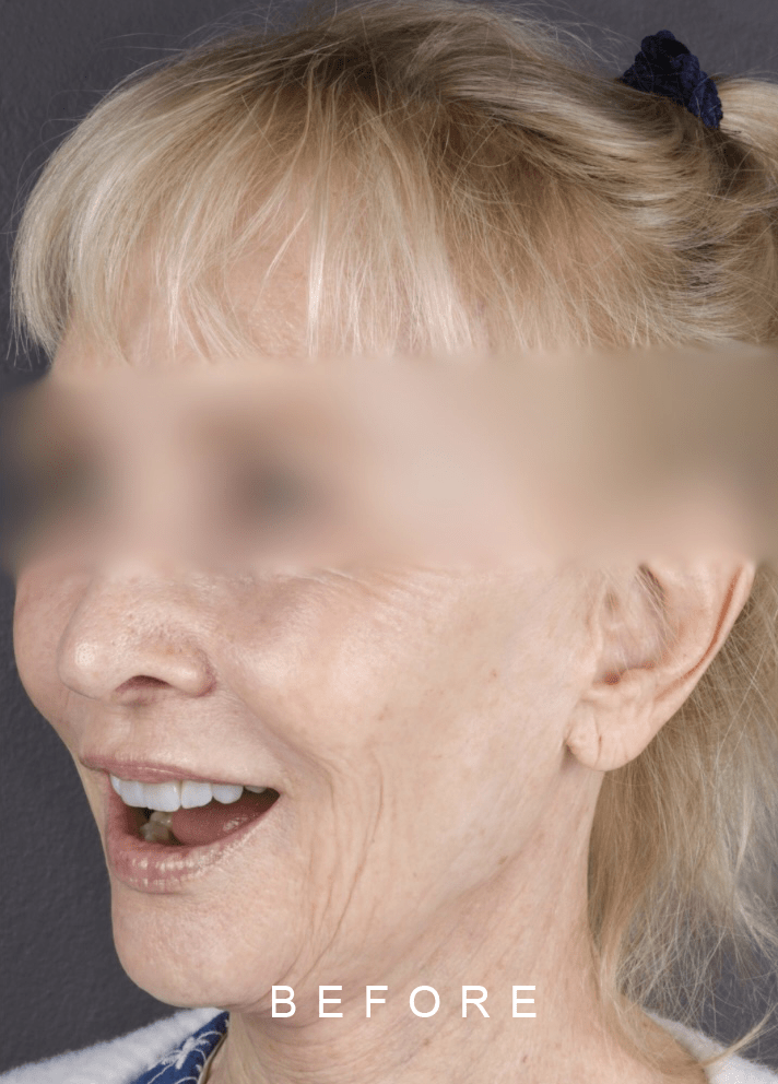
Figura 4: Fotos Extraorales sin tratamiento de ortodoncia / Extraoral Photos without orthodontic treatment
Figura 5. Fotos Extraorales frontal, lateral / Frontal and lateral extraoral photos
Figura 6: Fotos Intraorales / Intraoral Photos
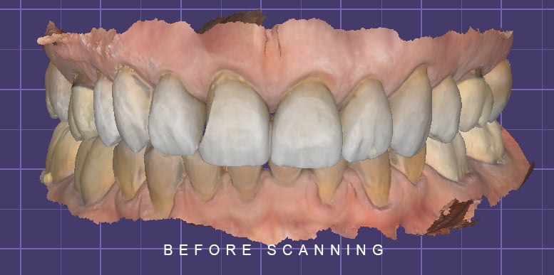
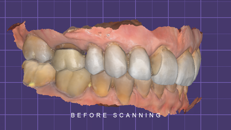
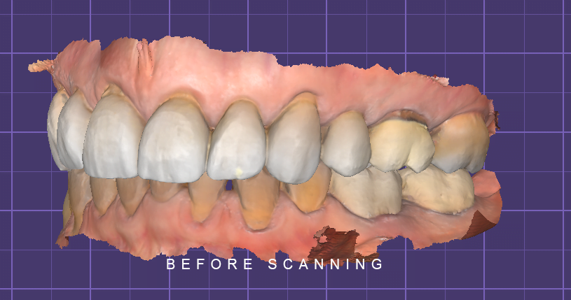
El plan de tratamiento lo ejecutamos por fases: Durante la primera fase, tomamos fotos extraorales e intraorales, toma de impresiones preliminares tanto manual como digital para planificación del caso (Figura.7).
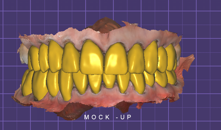
La segunda fase fue prueba de Mock-up con Protemp, color A1 marca 3M donde la aciente aprobó la forma, tamaño de los dientes. En cuanto al color; la paciente escogió el color BL3 MT de IVOCLAR VIVADENT.
La tercera fase fue el retiro de carillas, coronas dentales antiguas con fresa 4138F troncocónica halo verde, luego realizamos preparación dental con fresa 2134F torncocónica de diamante halo rojo, toma de impresiones definitivas en silicona de adición, impresiones manuales, digitales para la obtención de modelos previamente preparados y registro de mordida con era gris (Figura 8).
The treatment plan was divided through phases: First phase, we took extraoral and intraoral photos, preliminary impressions analogous and digital (Medit Link i700) for case planning (Figure.7).
The second phase was the Mock-up try-in with Protemp 4, color A1 from 3M where the patient approved the shape and size of the teeth. For the color, the patient chose the BL3 MT IVOCLAR VIVADENT.
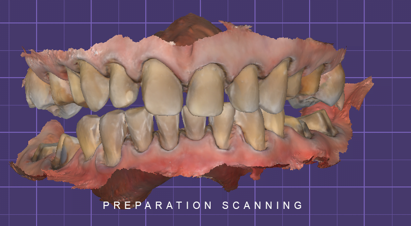
Third phase was the removal of veneers, old dental crowns with 4138F troncoconic green halo dental bur, then we prepared the teeth with 2134F torncoconic diamond red halo dental bur and took definitive impressions in addition silicone analogous and digital impressions to get prepared models and bite registration with gray wax (Figure 8).
Figura 7- Figure 7: escaneo dental sin preparación, mock -up digital , dental scanning without preparation, digital mock-up
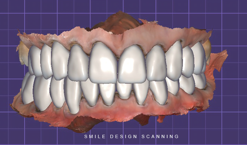
Figura 8. Fotos del escaneo de la preparación dental, confección de las carillas dentales en el programa Exocad / Scan of the dental preparation, digital smile design in the Exocad program
RESULTADO
Para la cuarta fase, comenzamos por el sector posterior. Realizamos prueba seca, húmeda para observar la adaptación de las coronas dentales , presencia de contactos prematuros y prueba del color.
Posteriormente, realizamos las respectivas pruebas de las carillas con glicerina, esperamos 5 minutos para que se hidraten, le mostramos a la paciente en luz natural cómo iba a lucir su sonrisa, sus dientes. Una vez aprobado por la paciente.
Realizamos la preparación tanto de las carillas y coronas como de la superficie dental y procedimos a la cementación individual de cada una.
Para las coronas dentales utilizamos Relyx Ultímate de 3M y para las carillas dentales cemento Choice 2 Bisco color translucent.
Retiramos excesos del cemento, selle palatino y ajuste de oclusión.
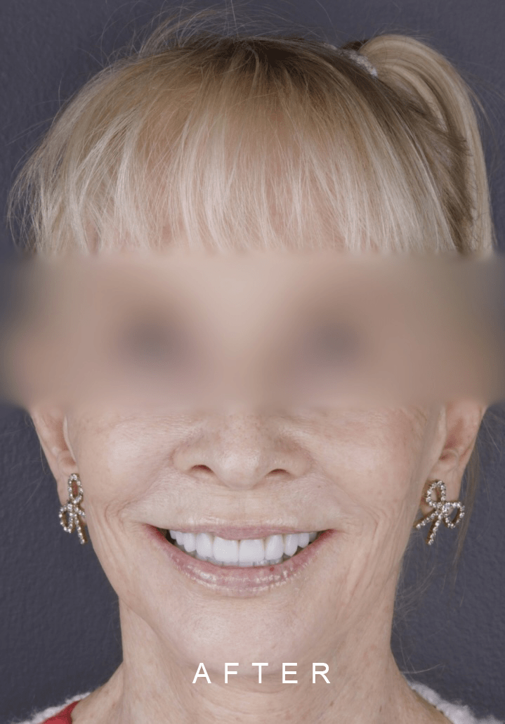
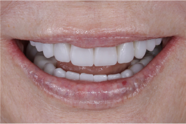
Figura 9. Resultado Final
RESULTS
For the fourth phase, we started with the molar area. We performed dry and wet test to observe the adaptation of the crowns, if there was any presence of premature contact and color test.
Subsequently, we performed the tests of the veneers with glycerin, waited for five minutes to hydrate them and we showed to the patient in natural light, how her smile and teeth would look like.
We prepared the veneers and crowns as well as the tooth surface and proceeded to the individual cementation of each one.
For the crowns we used Relyx Ultímate, translucent color from 3M and for the porcelain veneers System Choice 2 cement from BISCO, translucent color.
We removed excess cement, palatal seal and occlusal adjustment.
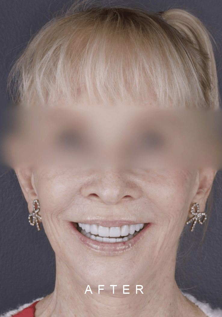
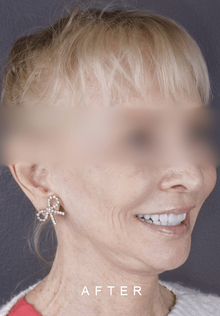
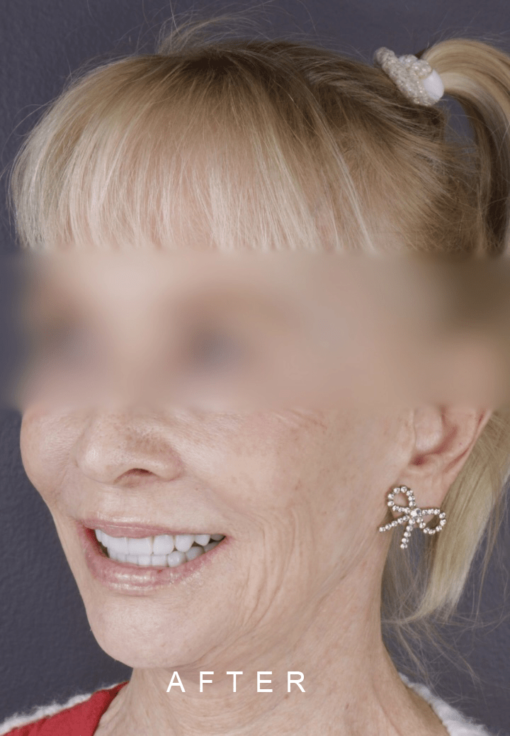
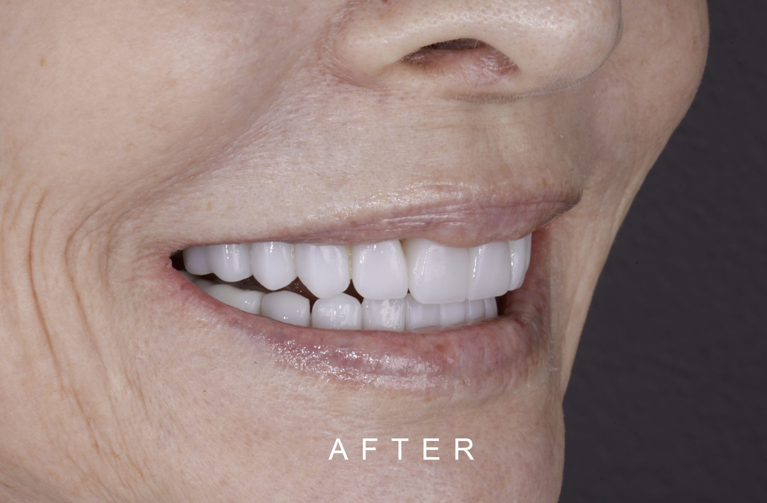
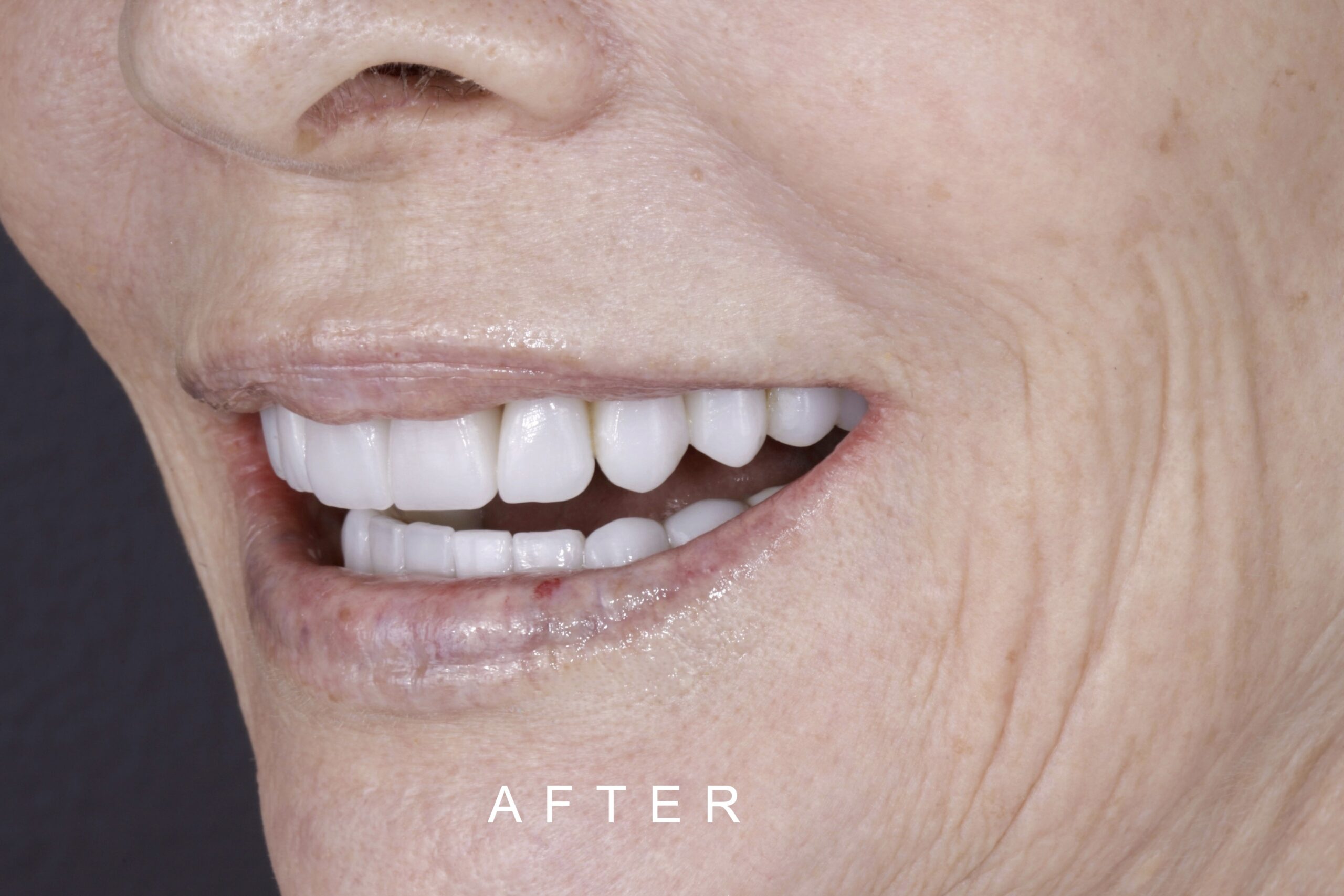
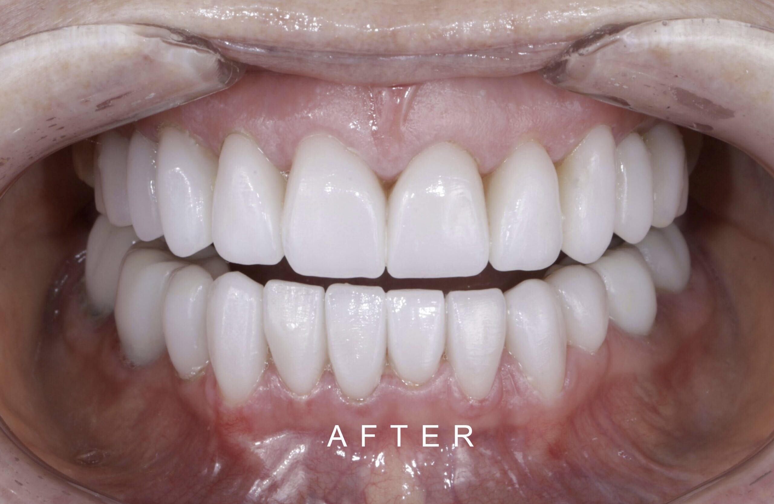
Figure 9. Final Result
AFTER FRONT FACE TOOTH WEAR 1
AFTER FRONT FACE TOOTH WEAR 2
AFTER FACE LATERAL TOOTH WEAR 1
AFTER FACE LATERAL TOOTH WEAR 2
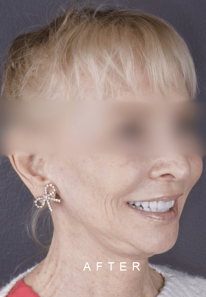
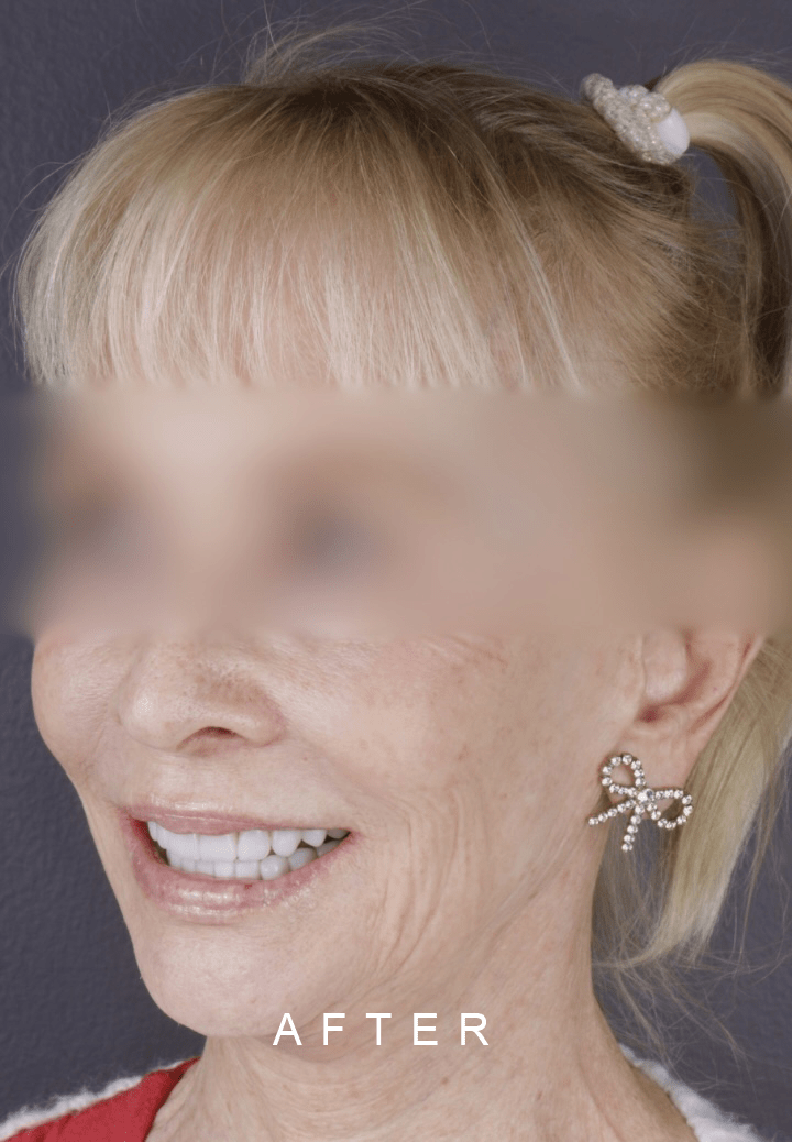
Figura 10. Fotos Extraorales (Después) / Extraoral Photos (After)
BEFORE FACE FRONT TOOTH WEAR 1
BEFORE FACE FRONT TOOTH WEAR 2
AFTER FRONT FACE TOOTH WEAR 1
BEFORE FACE LATERAL TOOTH WEAR 1
BEFORE FACE LATERAL TOOTH WEAR 2
AFTER FACE LATERAL TOOTH WEAR
BEFORE FACE LATERAL TOOTH WEAR
BEFORE FACE LATERAL TOOTH WEAR
AFTER FACE LATERAL TOOTH WEAR
Figura 12. Fotos extraorales antes y después / Extraoral before and after photos
BEFORE FRONT TOOTH WEAR 1
BEFORE FRONT TOOTH WEAR 2
AFTER FRONT TOOTH WEAR
BEFORE LATERAL TOOTH WEAR 1
BEFORE LATERAL TOOTH WEAR 2
AFTER LATERAL TOOTH WEAR
BEFORE LATERAL TOOTH WEAR
BEFORE LATERAL TOOTH WEAR
AFTER LATERAL TOOTH WEAR
BEFORE INTRAORAL FRONT TOOTH WEAR 1
AFTER INTRAORAL FRONT TOOTH WEAR 1
BEFORE INTRAORAL LATERAL TOOTH WEAR 2
AFTER INTRAORAL FRONT TOOTH WEAR 2
Figura 13. Fotos intraorales antes y después / Intraoral photos before and after
ENVEJECIMIENTO DENTAL
El envejecimiento de los dientes es el proceso de maduración y cambios que se dan en las piezas dentales y otros órganos de la boca. Estos se producen de manera natural, a medida que una persona se hace mayor. Al igual que el resto de los órganos del cuerpo humano, la boca sufre determinados cambios relacionados con el paso del tiempo y están muy relacionados con el cuidado oral que reciban a lo largo de la vida. Entre las condiciones que se pueden presentar:
Desgaste dental: es uno de los más comunes. Con el tiempo, el esmalte dental puede desgastarse debido al uso regular y al desgaste físico. Esto puede provocar que los dientes se vuelvan más cortos y pierdan la morfología, lo que a su vez puede afectar la mordida y la función masticatoria.
Cambios en el color: El esmalte dental también puede volverse más opaco y delgado con la edad, lo que puede hacer que los dientes luzcan más amarillos o manchados. Además, la acumulación de manchas de alimentos, bebidas y hábitos como fumar puede agravar este problema.
Recesión de las encías: Con el envejecimiento, es común que las encías se retraigan, exponiendo las raíces de los dientes. Esto puede causar sensibilidad dental y aumentar el riesgo de caries en las zonas expuestas.
Pérdida de dientes: La pérdida de dientes es más común en personas mayores debido a la enfermedad periodontal, la caries y otros factores relacionados con la edad. La ausencia dental puede tener un impacto significativo en la función de masticación y en la estética dental.
El envejecimiento dental, es un proceso que no se puede detener; por eso es importante tener una buena higiene bucal, visitar regularmente al odontólogo y adoptar hábitos saludables ya que nos va a garantizar saludl en todas las etapas de nuestras vidas.
TOOTH AGING
Tooth aging is the process of maturation and changes that happens in the teeth and other organs of the mouth. These happens naturally as a person gets older. Like the rest of the organs of the human body, the mouth undergoes certain changes related to the passage of time and are closely related to the oral care received throughout life. Among the conditions that may occur:
Dental wear: this is one of the most common. Over time, tooth enamel can wear down due to regular use and physical wear. This can cause teeth to become shorter and lose morphology, which in turn can affect bite and chewing function.
Changes in color: Tooth enamel can also become duller and thinner with age, which can cause teeth to appear more yellow or stained. In addition, the accumulation of stains from food, beverages and habits such as smoking can aggravate this problem. can aggravate this problem.
Receding gums: With aging, it is common for the gums to recede, exposing the roots of the teeth. This can cause tooth sensitivity and increase the risk of cavities in exposed areas.
Tooth loss: Tooth loss is more common in older people due to periodontal disease, tooth decay and other age-related factors. Missing teeth can have a significant impact on chewing function and dental esthetics.
Dental aging is a process that cannot be stopped; that is why it is important to have good oral hygiene, visit the dentist regularly and adopt healthy habits that will guarantee our health at all stages of our lives.